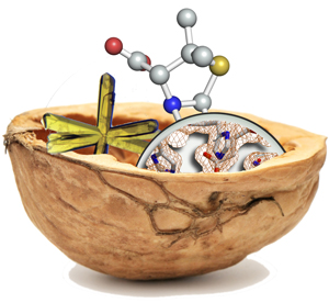
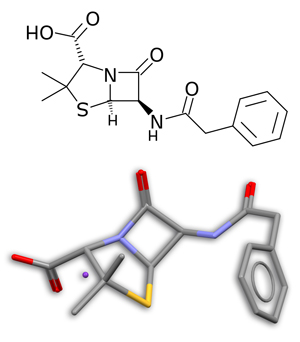
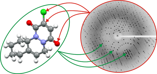
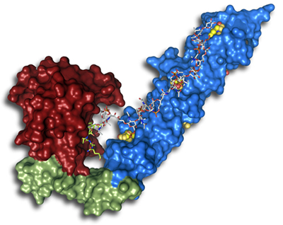
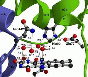
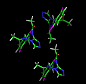
Crystallography in a nutshell
Mode: full-screen / central-screen / help
Table of contents through the logo
To the table of contents

If you don't have time or you are not keen to start reading all the information offered through the menu on the left (or in the table of contents), here you will find answers for a few questions that will help you to understand the beauty and capabilities of this part of science known as Crystallography:


What is Crystallography and what is it for?
 Up
 
By using crystals we can determine the atomic positions within matter. This can be applied to crystals made of either living or inanimate matter.

Crystallography is a branch of science that examines crystals. Today we know that crystals are made of matter, atoms, molecules and/or ions that fit together in repeating patterns, called unit cells, which like bricks stacked in three dimensions form the crystals. Inside the unit cells atoms are also repeated by symmetry operations. These patterns cause the crystals to show many sorts of unique shapes which for thousands of years have drawn our attention for their colors and outer beauty.

With the crystallographic tools developed during the 20th century, we can find out the inner structure of matter, living or inanimate, from which crystals are formed. To know the inner structure of matter means to determine the positions of all atoms and how they are linked together, in many cases forming atomic clusters known as molecules.
 
The atomic structure of matter generates knowledge that is used by chemists, physicists, biologists, and other scientists. This allows us not only to understand the properties of matter, but also to modify them for our benefit.

The two figures on the left show the molecular structure of penicillin, in the form of a diagram and its corresponding three-dimensional real shape. Only after the 3D molecular structure of penicillin was unambiguously determined in 1945 by Dorothy C. Hodgkin using crystallography, chemists could start the quick synthesis of this 
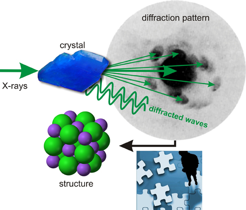
compound, thus saving millions of lives.

The importance of penicillin
Images taken from The National WWII Museum


Where does the word Crystallography comes from?
 Up

Crystal specimensThe word "crystallography" defines the science that "deals with the shapes and structures of crystals".

The word "crystal" comes from the Ancient Greeks 
κρνσταλλοσ (crustallos, or phonetically kroos'-tal-loscold + drop), who used the word crystal to refer to the mineralHexagonal close packing of oranges quartz, meaning both “ice” and “rock crystal”, that is cold and hard. Many minerals have always attracted our attention for its beautiful shapes and colors. There are references that relate Sumerians with the use of some mineral crystals in magic formulas, Chinese in its traditional medicine, and Egyptians that used crystals as jewels or in powder form for cosmetic purposes.

In its origins, Crystallography was dedicated exclusively to the study and description of the external appearance (morphology) of crystals, mainly minerals.

Over time we have found that crystals are ordered matter, ie, made up of atoms, ions and/or molecules perfectly stacked, as one can normally see pieces of fruit in a market. This packing is responsible for the striking forms that crystals normally show externally. 

Today Crystallography still deals with crystals, but its interest is mainly focused on their internal structure, not in their external shape.


When and how did crystallography begin as a science?
  Up

Although the first historical references to the use of crystals comes from the Ancient Sumerians (fourth millennium BC), it was not until the 17th and 18th centuries when the first scientific hypotheses about the inner nature of the crystals appeared, and all of them based on the observation of theirSnowflake external morphology.

Face angles and cellsThe German Johannes Kepler (1571-1630) was surprised by the fact that snowflakes landing on his coat always showed perfect six-cornered symmetry and never showed five of seven corners. He explained his observations in terms of the particle packings, as oranges show in the picture above.

 Also based on the observation of crystal morphologies, the Danish scientist Niels Stensen (1638-1686) and the French mineralogist Jean-Baptiste Louis Romé de l'Isle (1736-1790), established the law of constancy of angles between faces of different crystal specimens of the same species. With all this, René Just Haüy (1743-1822), another French mineralogist, commonly styled the Abbé Haüy, concluded that the crystals were made by the ordered stacking of small bricks, or unit cells, all of them identical.


Some years later, during the nineteenth century, the mathematical systematization of symmetry concepts governing the repetition of motifs around a point, as the petals of a flower around its axis, or the repetition of patterns by translation, gave rise to the establishment of some mathematical concepts such as:
In 1830, the German physician Johann Friedrich Christian Hessel (1796-1872), using the previous deductions of René Just Haüy (1743-1822), and after demonstrating that rotation axes can only be of orders 2, 3, 4 and 6, showed that the different possible morphologies of crystals can be combined to give exactly 32 different combinations of symmetry elements (the 32 point groups, also known as crystal classes).
Translations
In 1848 the French physicist Auguste Bravais (1811-1863) discovered that in the three-dimensional space, periodic repetitions by translation can only be made in 14 different modes (the so-called 14 Bravais lattices), as they must to be compatible with the 32 crystal classes.
Graphical representation of one of the 230 space groups

Finally, some 50 years later, the 14 Bravais lattices and the 32 crystal classes were the limitations that the Russian mathematician, crystallographer and mineralogist S. Evgraf Fedorov (1853-1919) and the German mathematician Arthur Moritz Schoenflies (1853-1928) used to independently deduce, between 1890 and 1891, the 230 space groups, which are the 230 possible ways to restrict distributions of repetitive structural units of the crystals (atoms, ions and molecules).

However, although these findings on the laws governing the structure of crystals were very important, they couldn´t help to solve the fundamental question: what is the size and shape of molecules inside the crystals? Or in short, what positions do atoms occupy in a crystal? Unfortunately, optical microscopes and visible light are not able to see such small pieces of matter as scientists imagined atoms to be!


The first big step forward: A new light to "look" inside crystals?
 Up

Illustration on the X-ray discovery
 The discovery of X-rays in 1895.
 (Illustration by Alejandro Martínez de Andrés, CSIC 2014)
 
The discovery of X-rays by Wilhelm Conrad Röntgen (1845-1923) in the late 19th century completely transformed the old field of Crystallography, which previously studied the morphology of crystals, mainly minerals. 

Although the discovery of this new "light" gave Röntgen a great popularity, it faded quickly. It took years until the utility of their amazing "light" was recognized as of medical interest, and even allowed him in 1901 to won the highest scientific award, the first Nobel Prize in Physics.


Thanks to that discovery Crystallography started to become one of the most important disciplines for many branches of science, especially for Mineralogy, Physics, Chemistry, Biology and Biomedicine. This was recently recognized by the United Nations, declaring 2014 International Year of Crystallography, and celebrating the centenary of the birth of modern Crystallography...


2014 International Year of Crystallography

To understand how Crystallography became an important branch of science, you should continue reading...


Laue experiment. X-rays are waves that diffract through the crystals
Laue showed in 1912 that X-rays are electromagnetic waves and that crystals have a regular internal structure.  

If Röntgen's discovery was important for the development of Crystallography, it was especially due to the experiment conducted in 1912 by Max von Laue (1879-1960), who convinced by his friend Paul Peter Ewald (1888-1985), decided to demonstrate the undulatory nature of X-rays and discovered the phenomenon of X-ray diffraction by crystals. He showed that X-rays are electromagnetic waves with a wavelength of about 10-10 meters and that the internal structure of crystals is regular, as if they had tiny slits of that order of magnitude, formed by the distances between atoms.

Indeed, after lighting a crystal of copper sulfate with X-rays Laue found that a photographic plate showed black spots not only at its center (as due to the fact that the incident beam passes directly through the crystal), but also in other places of the photographic plate, far away from the center. This result was interpreted as a consequence of the phenomenon called diffraction, whereby the X-ray beams scattered by the atoms interfere with each other inside the crystals and deviate from the central beam.


Experiment conducted in 1801 by Thomas Young, in an attempt to discern the wave or particle nature of light. Young investigated a pattern of interferences of light coming from a distant source and diffracted by two slits. This result contributed to the theory of the wave nature of light. Taken from Wikimedia


Representation of a wave

With this experiment, for which he received the Nobel Prize in Physics in 1914, Laue "killed two birds with one stone". Indeed, after watching the experimental results he could easily deduce that:
Regardless of the scientific interest of these two findings, readers who have arrived to this point will probably ask themselves:

Were Laue's results useful? This and other questions are answered below...


Crystals diffract X-rays. What is it useful for?
 Up

In 1914 Max von Laue (1879-1960) observed that X-rays are scattered in a peculiar way as they pass through crystals ... he discovered X-ray diffraction.


First diffraction pattern obtained by Max von Laue in 1914 using a crystal of copper sulfate and scheme on which the Bragg’s based their interpretation to deduce the internal structure of the crystals from the diffraction pattern.

It did not take long until the Max von Laue's discovery was recognized as very important. In fact, in the same year of Laue's experiment, William Henry Bragg (1862-1942) and his son William Lawrence Bragg (1890-1971) realized that if atoms inside crystals diffract X-rays and give rise to a diffraction pattern, this pattern should contain enough information to extract the relative positions of atoms in the crystal, that is to go backwards and retrace the path of diffraction. These scientists interpreted the phenomenon of diffraction with a simple geometric law: atoms in crystals occupy virtual planes that behave as mirrors, but reflecting X-rays only for certain angular positions of incident X-rays (Bragg's law). Father and son shared the Nobel Prize for Physics in 1915 for demonstrating the usefulness of the phenomenon discovered by von Laue in studying the internal structure of crystals.

The importance of Bragg's work cannot be overstated, for it heralded a revolution in the scientific understanding of crystals and their atomic arrangements. This discovery led to many of the most important scientific achievements of the last century, and these continue to the present day. To prove their theory, the Braggs were able to determine the atomic structure of simple materials such as sodium chloride (common salt) or the mineral zinc blende (zinc sulfide).

Although in those years these researchers were not able to solve the structure of more complex materials, over the time Crystallography has been able to answer a very large number of fundamental questions on various aspects of both inanimate and living matter.

 
Structural knowledge obtained by Crystallography allows us to produce materials with predesigned properties, from catalyst for a chemical reaction of industrial interest, up to toothpaste, vitro ceramic plates, extremely hard materials for surgery, or certain aircraft components, just to give some examples of small, or medium sized atomic or molecular materials.
 
Electronic materials... Lighter and stronger materials...
Digital signalling... Bone prostheses...
Electronics, sensitive screens... All kind of drugs and pharmaceuticals...
Electronics, high-resolution screens... Even improving foods, as chocolate...
 

Moreover, thanks to Crystallography we discovered the DNA secrets, the genetic code. We can increase the resistance of plants against environmental damage. We are able to understand, modify or inhibit enzymes involved in fundamental processes of life and important signaling mechanisms taking place inside of our cells, like cancer. With the structural knowledge of the ribosome, the largest factory of proteins in our cells, we can design new antibiotics and modify their structure to improve the efficiency. We are learning from the structure of certain components of some viruses to fight bacteria that are highly resistant to antibiotics, and are able to unravel the subtle defense machinery that has been developed from these microorganisms, so that we will be able to fight them with alternative tools.


 Atomic and molecular structure of a DNA fragment


How can atoms "be seen" with X-rays?
 Up

The interaction of X-rays with the atoms inside the crystals generates a kind of fingerprint known as diffraction pattern. Therefore, in order to "see" where the atoms are, we must be able to unravel the information contained in the diffraction pattern...


W.H. Bragg and W.L. Bragg’s findings opened unexpected ways to gain knowledge on the internal structure of matter. These two researchers were already aware that the right way to unravel the secrets of crystals was to go backwards, that is, stepping from the puzzle contained in diffraction pattern to the atomic positions in the crystal.

Although we don't claim to reveal all the details on how to solve that puzzle, we should give some idea about this supposed mystery...


To understand the difficulty we face, we need to remember what happens when a crystal is illuminated with X-rays...

When the X-ray waves pass through a crystal they interfere with each other, giving rise to waves that deviate from the line defined by the incident beam. When these new waves reach the photographic plate they produce a snapshot characteristic of each crystal species (such as a fingerprint). This is what we call a diffraction pattern. 


Example of how the two waves, shown at the top, add or subtract (depending on their relative positions), to generate a resultant wave (thick line below). Animation taken from The Pennsylvania State University
 
And although we cannot see them with our eyes, each of these waves is added to (or subtracted from) its neighbors, being reinforced or decreased, generating a resultant wave, as shown in the figure above. The resultant waves in each direction of diffraction are produced by the interferences occurring between the waves produced inside the crystal, a phenomenon entirely dependent on the relative positions of the atoms in the crystal.


The spots of the diffraction pattern are thus the result of the different waves arriving at the photographic plate. Each of these resultant waves is characterized by its amplitude (intensity) and a relative phase with respect to the remaining ones. In the figure below two resultant waves, each one with its own amplitude (intensity) and a relative phase, generate two separate spots in the diffraction pattern. The intensity of each spot is proportional to the amplitude of the corresponding wave, and each wave travels with a different relative phase.


At this point it seems therefore easy to deduce, at least qualitatively, that:
The intensities of the diffraction pattern (= the amplitudes of the waves) can easily be measured. However, there is no experimental procedure for measuring the relative phases of the waves!. Therefore, we can now understand why:
But, once the intensities of all diffracted waves are measured, if we are "somehow" able to find out their relative phases, there is a simple way (although with some numerical complexity) that allows us to determine the positions of all atoms in the crystal, that is, to calculate a three-dimensional picture of the inner structure of crystal. And this procedure applies for both living and inanimate matter.

Structure of a molecule inside a crystal
Representation of the three-dimensional structure of an organic molecule
 

Between the atomic positions in the crystal and the diffraction pattern there is a holistic relationship. The position of each atom in the crystal depends on the information existing in the diffraction pattern (wave intensities and relative phases). Conversely, the intensity and relative phase of each diffracted wave, shown as a spot in the pattern, depends on the position of all atoms in the crystal:


The position of each atom is determined by the sum of all diffracted waves (taking into account their intensities and relative phases). This kind of sum is a holistic process, ie, the information in every point of the crystal is dependent on the information existing in the whole diffraction pattern, and vice versa. It's the same thing as we can say of this arc of stones; the existence of the arc depends on each and every one of the stones...


Between the arc and the stones there is a holistic relationship. The arc depends on all and everyone of the stones.Image taken from definicionAB


Is this methodology similar to an optical microscope?
 Up

Although it has been said that determining the internal structure of crystals is not an immediate procedure, it resembles the process of watching a tiny object through an optical microscope. For example, to see the details of a fly wing (what we cannot distinguish directly with the naked eye), we put the wing on a slide, which is illuminated with visible light. Light passing through the wing is refracted producing various waves (with their intensities and relative phases) that go through a system of lenses that are able to "combine" these waves (with their intensities and phases) and produce an enlarged image showing the tiny details of the fly wing.

In what we might call the impossible X-ray microscope, the object being observed is a crystal that is illuminated with X-rays, instead of visible light. The waves of "light X” are diffracted by the crystal. But unfortunately, technically speaking there is no system of lenses able to "combine" diffracted waves of X-rays, with their amplitudes and phases, and therefore we have to be satisfied with the only thing we can measure on the diffraction pattern: the amplitudes (=intensities). 


Only after being able to calculate (somehow) the phases of the diffracted waves, we will be able to "see" the equivalent enlarged image of the optical microscope, that is, the internal structure of the crystal (=the atomic positions)...


The puzzle we are talking about refers to the calculation of the phases of the diffracted waves, but this is a problem that exceeds the limits of this summary...


Why do we use crystals instead of isolated molecules?
 Up

Some readers will probably wonder why we use crystals (packed molecules) to "see" atomic positions, rather than using isolated molecules...


The answer is quite simple. The interaction of X-rays with matter is very weak and if we irradiate an isolated molecule with X-rays, we get an interference pattern which contains very poor scattering information, making it difficult to reconstruct the structure of molecules from these interferences.


However, when a crystal is irradiated, the ordered molecular packing makes the crystal behave as a very powerful 'amplifier'. The cooperative interferences, called diffraction, produce a pattern containing much more information than the one created by an isolated molecule...


What kind of information is contained within a crystal structure?
 Up

The crystal structure contains full information, not only about the spatial positions occupied by all atoms, but also on their thermal vibrational states. From these positions we can also derive if atoms are bound forming molecules, and easily calculate all type of geometrical parameters, including bond lengths, bond angles, etc.  In addition, the structural crystallographic data also show very rich information about how molecules interact with each other, through attractions or repulsions.

Crystallography is even able to distinguish between a molecule and its mirror image (see left image). These molecules seem to be the same, but they are quite different, and so are their properties. Think for instance on our two hands; they seem to be "equal", but in reality they are very different; they are not superimposable, they are mirror images.

Molecules related by a mirror plane
Two mirror-image molecules

The three-dimensional shape of a molecule relates to how the molecule will work in a chemical reaction, in a test tube, or inside a living being. Once the relationship between the structure and properties is understood, it is often possible to modify or design new materials or molecules with specific desired properties.

Today, crystallographers study the atomic structure of any material that can build a crystal, from very simple substances to viruses, proteins or huge protein complexes. But they also investigate a wide variety of other materials, such as membranes, liquid crystals, fibers, glasses, liquids, gases and quasicrystals. In short, the atomic and/or molecular structure that Crystallography provides contains the most relevant information to understand the properties of materials, so we can modify them to our benefit. Moreover, current macromolecular crystallography includes new X-ray sources, enhanced remote-accessible capabilities and time-resolved methods to capture intermediate structures along reaction pathways.


Crystallography allows us to know all geometrical details (interatomic distances, bond angles, etc.) of molecules, even for huge protein complexes


Crystallography provides full details on both molecular packing and intermolecular interactions.


The structures of most known materials (inorganic, organic, metal-organic, proteins, enzymes, nucleic acids...) are available through specialized databases that are continuously updated. 

Even if you do not pretend to know anything more about crystallography, we hope this summary has clarified some ideas about this powerful and beautiful branch of modern science.


Table of contents
Introduction